Have you ever wondered about the intricate details of a tiny insect’s wing, the mesmerizing patterns on a pollen grain, or the fascinating structure of a single blood cell? The world around us is teeming with wonders invisible to the naked eye, waiting to be explored. It’s the microscope, a marvel of scientific ingenuity, that unlocks this hidden realm. This powerful tool, with its intricate parts and finely tuned mechanics, allows us to magnify the unseen and unravel the mysteries of the microscopic world.

Image: microscopeinternational.com
But how does it work? What are the essential components that come together to reveal the beauty and complexity of the miniature universe? This article will take you on an exciting journey, diving deep into the parts of a microscope and understanding their crucial functions. Prepare to have your curiosity piqued as we unravel the magic behind this indispensable tool, empowering you to see the world in a whole new light.
A Glimpse into History: From Simple Lenses to Modern Microscopes
The journey of the microscope dates back centuries. The invention of the magnifying glass in the 13th century paved the way for early lens-based observation. Zacharias Janssen, a Dutch spectacle maker, is often credited with building the first compound microscope in the late 16th century. This device used two lenses to magnify objects further than single lens instruments, marking a significant leap in scientific exploration.
However, it wasn’t until the 17th century that the microscope truly began to revolutionize scientific understanding. Antonie van Leeuwenhoek, a Dutch merchant and scientist, perfected lens grinding techniques, achieving magnifications of up to 200x. His meticulous observations of microscopic organisms, including bacteria, protozoa, and red blood cells, opened a new world of microscopic life, forever changing how we viewed the natural world.
The Foundation of Clarity: The Structure of a Compound Microscope
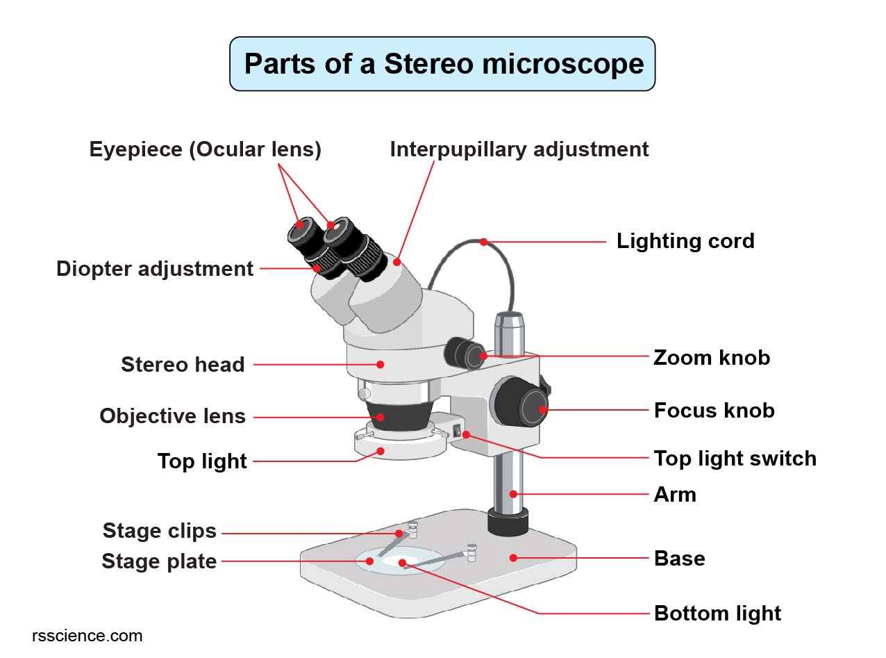
A compound microscope, the most common type found in educational and research settings, is a complex yet elegantly designed instrument. Its core components work harmoniously, each playing a vital role in creating a magnified image for observation. Here’s a breakdown of the key parts and their functions:
1. The Base: The foundation of the microscope, providing stability and support for the entire structure.
2. The Arm: The vertical structure connecting the base and the stage, allowing for the adjustment and movement of the microscope.
3. The Stage: A flat platform where the specimen is placed for observation. It often includes clips to hold the specimen in place.
4. The Light Source: A built-in illumination system that projects light onto the specimen. This can be a built-in LED, halogen lamp, or a mirror reflecting external light.
5. The Condenser: A lens positioned below the stage to focus the light beam onto the specimen. This ensures even illumination across the sample.
6. The Diaphragm: A mechanism for controlling the amount of light passing through the specimen. Adjusting the diaphragm allows for optimal contrast and clarity in the image.
7. The Objective Lenses: A set of lenses mounted on a revolving nosepiece, providing different levels of magnification. The typical objective lens magnifications include 4x, 10x, 40x, and 100x (oil immersion).
8. The Revolving Nosepiece: A rotating mechanism that holds the objective lenses and allows for easy switching between different magnifications.
9. The Body Tube: The vertical cylinder connecting the objective lenses to the eyepiece, providing the pathway for light to travel from the objective to the eye.
10. The Eyepiece: A lens at the top of the body tube that magnifies the image projected by the objective lens. The eyepiece typically has a magnification of 10x.
11. The Fine Adjustment Knob: A precise knob used for fine focusing, creating sharp images at high magnification.
12. The Coarse Adjustment Knob: A larger knob for initial focusing, bringing the specimen into view quickly.
13. The Mechanical Stage Control Knobs: These knobs allow for precise movement of the specimen horizontally and vertically on the stage.
The Magic of Magnification: How a Microscope Forms an Image
The process of forming an image using a compound microscope involves a fascinating interplay of light and lenses. This journey begins with the light source, shining light through the condenser, focusing it onto the specimen. The specimen, placed on the stage, becomes illuminated, allowing details to be visible.
As light passes through the specimen, it enters the objective lens, the first lens system. This lens magnifies the specimen and forms a real, inverted image. This magnified image is then projected up the body tube, passing through the eyepiece, the final lens system. The eyepiece further magnifies the image, producing a virtual, magnified, and upright image that the observer sees.

Image: www.vrogue.co
Exploring the World of Microscopy: Applications and Innovations
The microscope is not just a tool for scientific curiosity; it’s a fundamental instrument used in various fields, from medicine to engineering:
1. Biomedical Research: Microscopes are vital in diagnosing diseases, studying cell structure, and observing bacteria, viruses, and other microscopic organisms.
2. Material Sciences: Microscopy helps examine material properties, surface characteristics, and defects in various materials, leading to advancements in manufacturing, engineering, and nanotechnology.
3. Quality Control: In industries ranging from pharmaceuticals to food processing, microscopes ensure product quality, detect impurities, and guarantee safety.
4. Environmental Science: Microscopes are used for analyzing water and soil samples, identifying pollutants, and understanding the microscopic organisms that form the basis of ecosystems.
5. Forensic Science: Microscopes are essential in crime investigations, analyzing fibers, hair, and other microscopic evidence to identify suspects and build strong cases.
Beyond Traditional Microscopes: Exploring New Frontiers
The field of microscopy continues to evolve, with new technologies emerging to push the boundaries of observation.
1. Electron Microscopy (EM): Using beams of electrons instead of light, EM achieves incredible resolutions, allowing for the visualization of subcellular structures like organelles and even individual atoms.
2. Scanning Probe Microscopy (SPM): This technique employs a fine probe to scan the surface of a specimen, providing a detailed topographical map of the sample at the atomic level.
3. Confocal Microscopy: This advanced technique utilizes laser light and optical sections to create high-resolution images of thick specimens, allowing researchers to study 3D structures with incredible detail.
These innovations are opening up new avenues for scientific understanding, enabling researchers to study the world at a level of detail never before achieved.
Embracing the Microscopic World: Tips for Enhancing Your Observations
Here are some tips you can use to get the most out of your microscope and enhance your observations:
1. Cleanliness is Key: Before using your microscope, always clean the lenses and stage with lens paper to remove dust or smudges that can distort your image.
2. Proper Illumination: Adjust the diaphragm and condenser to achieve optimal lighting. Too much light may wash out details, while too little light may make the specimen appear blurry.
3. Starting with Low Magnification: Begin with the lowest magnification objective lens and gradually increase as needed. This allows you to find the specimen easily and then focus in on the details at higher magnifications.
4. Patience and Practice: Using a microscope effectively requires practice. Experiment with different specimens, lighting techniques, and magnifications to discover the optimal settings for your observations.
5. Documentation: Record your observations with detailed descriptions, sketches, or even photographs to document your discoveries and ensure accurate reporting.
Parts Of Microscope And Functions Pdf
A World of Discovery Awaits
The microscope has opened a window into a world that was previously hidden from view. By understanding the functions of its various parts, you can explore the intricate details of the microscopic world, unlocking the secrets of nature and pushing the boundaries of scientific exploration. So, grab your microscope, embrace your curiosity, and embark on your own journey of scientific discovery.


